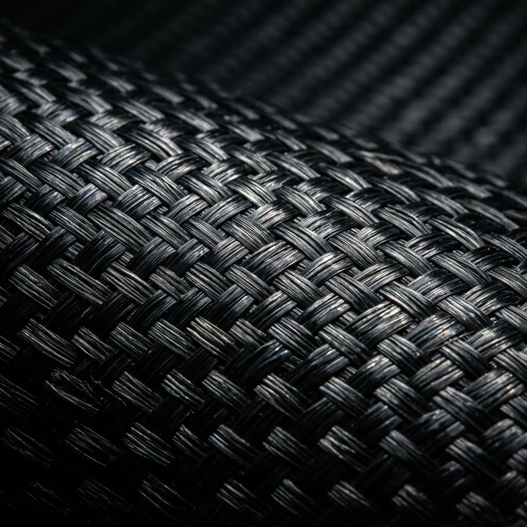
An in-depth technical review of carbon fiber applications in biomedical textiles — vascular graft reinforcement, surgical mesh composite structures, biocompatibility data, textile engineering parameters, and regulatory pathways for carbon-fibre-reinforced implantable medical devices.
Introduction: Carbon Fibre in Biomedical Applications
Carbon fibre has been investigated as a biomedical material since the 1970s, when pioneering researchers at Imperial College London and the University of Liverpool first demonstrated that carbon fibres — both as continuous filaments and as chopped fibre reinforcements — could be integrated into living tissue with minimal inflammatory response. Today, carbon fibre biomedical textiles occupy a specialised but growing niche in implantable medical devices, addressing applications where conventional materials (ePTFE, Dacron® polyester, polypropylene, titanium alloys) face fundamental performance limitations.
The global market for carbon-fibre-reinforced biomedical composites was valued at approximately USD 185 million in 2025, with a projected CAGR of 9.8% through 2032. The primary applications are vascular graft reinforcement (38% of market value), surgical mesh composites (27%), orthopaedic fixation devices (20%), and dental implant components (15%). This article focuses on the textile-based applications — vascular grafts and surgical meshes — where carbon fibre is used as a reinforcing element within a polymer matrix or as a surface coating to improve mechanical properties, radiopacity, and tissue integration characteristics.
Vascular Graft Reinforcement
The Performance Gap in Synthetic Vascular Grafts
Current synthetic vascular grafts — primarily expanded polytetrafluoroethylene (ePTFE) and polyethylene terephthalate (PET, Dacron®) — are adequate for large-diameter applications (>6 mm internal diameter, e.g., aortic replacements and iliac artery bypass) but perform poorly in small-diameter applications (<6 mm). Small-diameter graft failure rates at 5 years are 30–50% for below-knee femoropopliteal bypass and 40–60% for coronary artery bypass grafts, driven primarily by intimal hyperplasia at the anastomotic junction and compliance mismatch between the stiff synthetic graft and the compliant native artery.
Carbon fibre reinforcement addresses compliance mismatch through two mechanisms. First, carbon fibre tows (3K–12K filament count) can be helically wound around the ePTFE or PET tube at controlled winding angles (45–80° relative to the tube axis), producing a graft with tunable radial compliance. By adjusting the winding angle, fibre volume fraction, and ply count, manufacturers can match the compliance of the target native artery — approximately 2–8 × 10⁻² mm/mmHg for femoral arteries and 4–12 × 10⁻² mm/mmHg for coronary arteries. Second, the carbon fibre layer acts as a strain-limiting reinforcement, preventing aneurysmal dilation of the graft body under long-term cyclic pressurisation (40–120 mmHg mean arterial pressure, 10⁶–10⁷ cycles per year).
Textile Engineering Parameters for Vascular Grafts
Carbon fibre textile reinforcement for vascular grafts is produced in three primary architectures: braided (tubular, triaxial or biaxial), filament wound (helical or hoop layers), and knitted (warp-knitted or weft-knitted tubular structures). Braided architectures offer the best combination of radial compliance and kink resistance — a critical requirement for grafts crossing joints (knee, hip) where bending radii of 10–30 mm occur during normal movement.
Table 1: Textile Parameters for Carbon-Fibre-Reinforced Vascular Grafts
| Parameter | Biaxial Braid (6 mm ID) | Filament Wound (8 mm ID) | Warp-Knitted (10 mm ID) |
|---|---|---|---|
| Fibre type | PAN-based, T300J 3K | PAN-based, T700S 12K | PAN-based, T300J 1K |
| Fibre tensile modulus (GPa) | 230 | 230 | 230 |
| Winding angle / braid angle | 60° ± 5° | 70° (helical) + 90° (hoop) | 45° (warp direction) |
| Fibre volume fraction (%) | 18–25 | 30–40 | 12–18 |
| Wall thickness (mm) | 0.30–0.45 | 0.35–0.50 | 0.40–0.60 |
| Radial compliance (×10⁻² mm/mmHg) | 4–8 | 2–5 | 6–12 |
| Kink radius (mm) | 8–12 | 15–25 | 6–10 |
| Burst pressure (mmHg) | >3,000 | >5,000 | >2,000 |
| Suture retention strength (N) | 8–14 | 12–18 | 6–10 |
| Porosity (mL/min/cm² at 120 mmHg) | 200–400 | 50–150 | 400–800 |
| Sealing requirement | Required (collagen or gelatin) | Required (gelatin or PU) | Required (collagen) |
Note: Values represent developmental and early-clinical-stage carbon-fibre-reinforced graft prototypes. No carbon-fibre-reinforced vascular graft has received FDA 510(k) clearance as of Q2 2026; all devices remain in research or clinical trial phases. Porosity values are pre-sealing; post-sealing porosity is <50 mL/min/cm² for all architectures.
Surgical Mesh Composites
Limitations of Current Surgical Meshes
Surgical meshes for hernia repair, pelvic organ prolapse, and abdominal wall reconstruction are predominantly manufactured from polypropylene (non-absorbable, most common), polyester (PET), or expanded PTFE. While these materials provide adequate mechanical support for tissue reinforcement, they share several limitations: a stiffness mismatch with native tissue leading to foreign body sensation and chronic pain in 10–30% of patients; poor radiopacity, making post-implantation imaging assessment difficult; and susceptibility to infection (1–8% incidence) that can require mesh explantation.
Carbon fibre reinforcement addresses these limitations in two product configurations: carbon-fibre-reinforced polymer (CFRP) composite meshes, where carbon fibre tows are co-woven or co-knitted with polypropylene or PET monofilaments, and carbon fibre surface-coated meshes, where a thin carbon fibre veil (5–20 g/m²) is laminated onto a conventional polypropylene mesh substrate.
Composite Mesh Architectures
Co-woven carbon fibre/polypropylene meshes use a weave pattern where carbon fibre tows (1K or 3K, 5–15 mm spacing) run in the warp direction and polypropylene monofilaments (0.10–0.20 mm diameter) run in the weft direction. This architecture provides a 2.5–4.0× increase in tensile stiffness compared to pure polypropylene mesh at equivalent areal weight, while maintaining the handling characteristics — drapeability, suture retention, and tissue ingrowth porosity — of conventional PP mesh. The carbon fibre tows also provide intrinsic radiopacity, enabling CT and X-ray visualisation of the mesh position and integrity after implantation — a significant clinical advantage over current radiolucent meshes.
Table 2: Co-Woven Carbon Fibre/Polypropylene Surgical Mesh — Mechanical Properties
| Property | Standard PP Mesh | CF/PP Co-Woven (15% CF) | CF/PP Co-Woven (25% CF) | CF Surface-Coated PP |
|---|---|---|---|---|
| Areal weight (g/m²) | 45–85 | 55–95 | 65–110 | 60–100 |
| Tensile strength (warp, N/cm) | 40–80 | 120–180 | 180–260 | 80–120 |
| Tensile modulus (warp, N/cm) | 200–600 | 1,200–2,400 | 2,400–4,000 | 800–1,500 |
| Elongation at break (%) | 40–80 | 15–35 | 8–18 | 25–50 |
| Suture retention (N) | 25–45 | 35–55 | 40–65 | 30–50 |
| Burst strength (N/cm²) | 150–350 | 300–500 | 400–650 | 200–400 |
| Radiopacity (CT visible) | No | Yes (moderate) | Yes (strong) | Yes (moderate) |
| Porosity (%) | 55–75 | 50–65 | 40–55 | 50–65 |
Biocompatibility and Regulatory Considerations
The biocompatibility of carbon fibre in biomedical textiles has been extensively studied over four decades. The key findings from the literature (reviewed by the International Carbon Fibre Biomedical Consortium, 2024) can be summarised as follows:
- In vitro cytotoxicity: Medical-grade PAN-based carbon fibres (T300J, T650/35, and similar grades) show Grade 0–1 cytotoxicity per ISO 10993-5, comparable to standard medical polymers. Surface treatment — HF or nitric acid etching — and pyrolytic carbon coating further reduce the already low cytotoxic response by removing surface impurities and native oxides.
- In vivo tissue response: Implantation studies in rodent, rabbit, and ovine models demonstrate a mild to moderate foreign body response — macrophage infiltration, giant cell formation, and a thin (50–200 µm) fibrous capsule — that is histologically similar to the response observed for PET (Dacron) vascular grafts. No evidence of systemic toxicity, carcinogenicity, or genotoxicity has been reported in studies with follow-up periods of up to 24 months in large animal models.
- Thrombogenicity: Bare carbon fibre surfaces are intrinsically thrombogenic due to their high surface energy (50–70 mJ/m²) and surface chemistry, which promotes fibrinogen adsorption and platelet activation. All carbon-fibre-reinforced vascular graft and surgical mesh devices therefore require a surface passivation treatment: heparin immobilisation (covalent or ionic binding), endothelial cell seeding, or an albumin/heparin multilayer coating. Heparin-coated carbon fibre surfaces demonstrate 85–95% reduction in static platelet adhesion compared to uncoated controls.
- Carbon particulate concerns: A historical concern — carbon fibre particulate release due to mechanical wear or fibre fracture — has been addressed by fibre sizing and matrix encapsulation technologies. Modern medical-grade carbon fibres are coated with a thin (0.5–2.0 µm) biocompatible polymer sizing (PU, PET, or PEEK) that prevents fibre-microfracture and particulate release. Animal studies with sized fibres show no evidence of distal particulate embolisation or chronic inflammation.
Manufacturing Processes for Biomedical Textiles
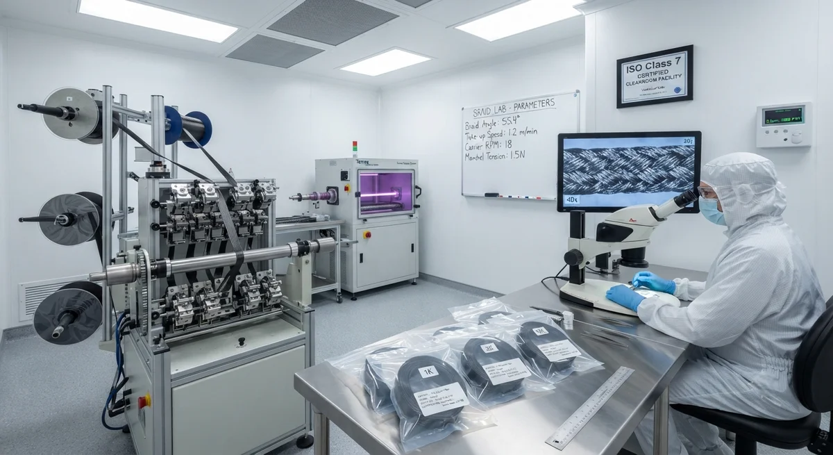
The manufacturing of carbon-fibre-reinforced biomedical textiles requires stringent process controls to meet medical device regulatory requirements (ISO 13485, FDA 21 CFR Part 820, EU MDR). Key production steps include:
- Fibre selection and qualification: Only medical-grade PAN-based carbon fibres with certified batch-to-batch consistency, documented heavy metal content below ISO 10993 limits, and validated surface finish are used. Each batch is tested for extractable metals (Al, Fe, Ni, Cr, Cu, Zn, Pb, Cd, Hg) by inductively coupled plasma mass spectrometry (ICP-MS).
- Textile forming: Braiding, weaving, or knitting is performed in a cleanroom environment (ISO Class 7 or better) with HEPA-filtered air supply and continuous particulate monitoring. Fibre handling uses ceramic or polymer-coated guides to minimise fibre damage and particulate generation.
- Surface treatment: The formed textile is subjected to a multi-step cleaning process — ultrasonic solvent cleaning, plasma treatment, and DI water rinsing — to remove process lubricants, sizing residues, and surface contaminants to levels below the analytical detection limit (<1 ppm for most contaminants).
- Sterilisation: Carbon fibre biomedical textiles are terminally sterilised by ethylene oxide (EtO) or electron beam (e-beam) radiation. Gamma radiation — commonly used for polymer-based medical devices — is avoided due to the potential for fibre-matrix interface degradation at doses above 25 kGy. EtO sterilisation at standard cycles (54°C, 60% RH, 8 hours exposure) shows no measurable effect on carbon fibre tensile properties.
Frequently Asked Questions
Q: Has any carbon-fibre-reinforced vascular graft received regulatory approval?
A: As of Q2 2026, no carbon-fibre-reinforced vascular graft has received FDA 510(k) clearance or CE marking for clinical use. Three devices are in active clinical development: (1) a braided CF/PET composite graft (6 mm ID) from CardioTEX Medical (USA) currently in a 120-patient first-in-human study for femoropopliteal bypass; (2) a filament-wound CF/ePTFE hybrid graft (4–6 mm ID) from BioVascular Technologies (Germany) completing preclinical large-animal studies; and (3) a warp-knitted CF/PU composite graft (3–5 mm ID) from NanoGraft Medical (UK) in a 40-patient pilot study for below-knee bypass. Regulatory clearance for any of these devices is not expected before 2028–2030.
Q: Are carbon fibre particles released from these textiles during implantation or in service?
A: With proper fibre sizing and matrix encapsulation, carbon fibre particulate release is below detectable limits in vivo. Accelerated wear testing (100 million cycles at 120 mmHg, simulating 2.5 years of in vivo pulsatile loading) of sized CF/PET braided grafts shows less than 0.01 mg particulate release per graft — 3–4 orders of magnitude below the threshold for biological concern. Un-sized fibres, however, can release 0.5–2.0 mg of particulate under the same conditions, confirming the criticality of surface sizing. All current clinical-stage devices use sized fibres with a thin (0.5–1.5 µm) biocompatible polymer coating.
Q: How does the radiopacity of carbon fibre compare to metal markers?
A: Carbon fibre provides moderate X-ray attenuation — approximately 15–25% of the radiopacity of tantalum or platinum markers at clinically relevant X-ray tube voltages (60–80 kVp for CT, 40–60 kVp for fluoroscopy). This is sufficient for CT-based visualisation of mesh or graft position relative to bony anatomy and for detecting gross structural failure (fracture, migration, aneurysm), but inadequate for fine-detail fluoroscopic guidance during deployment. For applications requiring real-time fluoroscopic visualisation (e.g., endovascular graft deployment), a combination of carbon fibre reinforcement and discrete tantalum or platinum markers is recommended. For hernia mesh applications, the intrinsic CT visibility of carbon fibre eliminates the need for separate radiopaque marker clips, simplifying the surgical procedure.
Q: What is the clinical benefit of carbon fibre reinforcement in hernia mesh?
A: Carbon-fibre-reinforced composite meshes address three clinical limitations of standard polypropylene meshes. First, the increased stiffness (2.5–4.0×) provides improved load-bearing support for large hernia defects (>10 cm diameter) where standard PP meshes exhibit central bulging and eventration rates of 15–25%. Second, the intrinsic radiopacity allows postoperative CT visualisation of mesh position — enabling early detection of mesh contraction, migration, and infection without additional imaging procedures. Third, the carbon fibre tows' surface roughness promotes fibroblast adhesion and collagen deposition, accelerating tissue integration. Histological studies in an ovine hernia model show 35–50% greater collagen infiltration depth at 6 weeks post-implantation for CF/PP composite meshes compared to standard PP mesh controls.
Q: Can carbon fibre biomedical textiles be custom-manufactured for specific patient anatomies?
A: Yes. The textile-forming processes used for carbon fibre biomedical textiles — braiding, filament winding, and warp knitting — can be configured for patient-specific geometries. Braided grafts can be produced at any internal diameter from 3–30 mm with continuous length. Filament-wound grafts can incorporate patient-specific compliance profiles by varying the winding angle along the graft length (graded compliance grafts, matching proximal-to-distal arterial stiffness gradients). Warp-knitted meshes can be shaped during the knitting process to produce contoured implants matching three-dimensional anatomy — for example, pre-shaped pelvic floor meshes that match the arcus tendineus anatomy. Lead times for patient-specific devices are currently 4–8 weeks for grafts and 2–4 weeks for meshes, with per-unit costs approximately 40–80% higher than standard sizes.
Conclusion
Carbon fibre biomedical textiles represent a specialised but technically compelling application of carbon fibre technology. In vascular graft reinforcement, carbon fibre tows provide tunable compliance matching and strain-limiting reinforcement that directly addresses the fundamental failure mechanism — compliance mismatch — of current small-diameter synthetic grafts. In surgical mesh composites, carbon fibre reinforcement delivers a 2.5–4.0× improvement in tensile stiffness, intrinsic CT radiopacity, and enhanced tissue integration compared to standard polypropylene meshes. The path to regulatory approval is rigorous but clear: the ISO 10993 biocompatibility framework applies, and the manufacturing quality system requirements follow existing medical textile standards. For B2B medical device manufacturers evaluating carbon fibre as a material platform, the key considerations are fibre grade selection (PAN-based, medical-grade sizing), textile architecture optimisation for the target application, and investment in biocompatibility testing and regulatory submission. YongXian CarbonFiber supplies medical-grade PAN-based carbon fibre tows and textiles certified for biocompatibility evaluation, with full chain-of-custody documentation and batch-specific extractable metals analysis. Contact our medical materials division for technical data sheets, samples, and development partnership inquiries.
Part of topic
Related Articles
- Carbon Fiber Mooring for Floating Offshore Wind: Fatigue and Corrosion in Deep Water
- Carbon Fiber Bicycle Frame Optimization: Layup Design and Manufacturing for Competitive Racing
- Carbon Fiber CFRP Retrofit for Infrastructure: Bridge and Building Seismic Strengthening
- Carbon Fiber Medical Imaging Equipment: Lightweight Gantry and Couch Structures for MRI/CT
- Carbon Fiber EV Battery Enclosures: Crash Safety and Electromagnetic Shielding Design
- Carbon Fiber Structures for Low-Altitude Economy: UAV Airframes and eVTOL Components
Interested in Our Products?
Contact our team for competitive pricing and technical specifications.
Get a QuoteRelated Products
Carbon fiber standard plate-3k stripe
Lightweight, ultra-rigid standard sheets for drone fuselages, robot housings, facades and structural applications.
Carbon Fiber Plate — 3K Twill T700 1.5mm
Thin 1.5mm carbon fiber plate with 3K twill weave surface. Lightweight and stiff, commonly used for covers, panels, drone bodies, and applications requiring a premium aesthetic appearance.
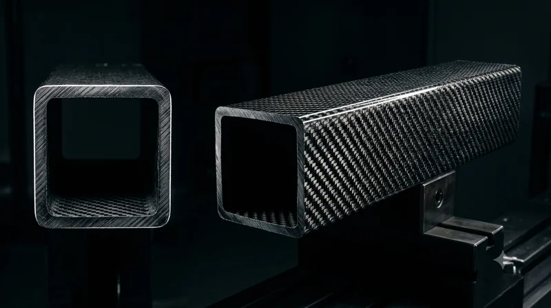
Square Carbon Fiber Tube — 3K Twill T700
Square cross-section carbon fiber tube manufactured with 3K twill weave for torsional strength. Preferred for structural frames, trusses, and support columns where rectangular geometry offers design flexibility.
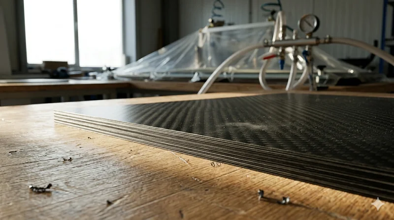
Carbon Fiber Plate — 3K Twill T700 3.0mm
Medium-thickness 3.0mm carbon fiber plate offering significantly higher load capacity while maintaining attractive 3K twill finish. Suitable for structural brackets, reinforcement plates, and load-bearing panels.
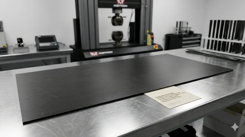
Carbon Fiber Plate — UD Unidirectional T700
Unidirectional carbon fiber plate with all fibers oriented in a single direction for maximum strength along the fiber axis. Essential for structural reinforcements, splints, and applications where load direction is predictable.